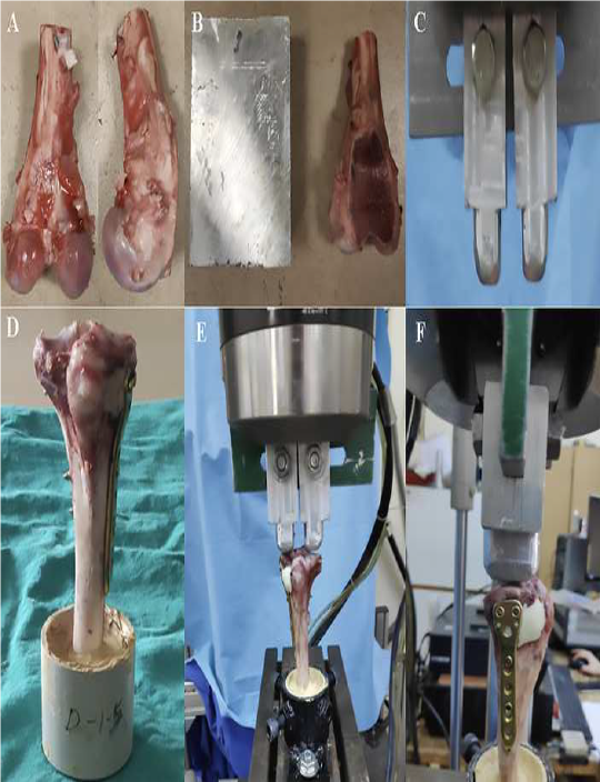
In this study, which was carried out jointly by ITU, Faculty of Mechanical Engineering, Biomechanics and Strength Laboratory and Istanbul Basaksehir Pine and Sakura City Hospital, Orthopedics and Traumatology Clinic, the cementation and internal fixation methods applied to bone defects of varying sizes after proximal tibial resection were biomechanically compared and the biomechanical method of the ideal fixation method according to the defect size was determined. It is intended to be explained by tests. Cement filling and plate fixation groups were found to have the highest failure load in all defect sizes. Cement+screw combination below 25% defect size reduces the risk of tibial plateau and metaphyseal fractures. It was also determined that fracture complexity was significantly related to defect size and fixation method. The study made a significant contribution to the literature. Because the distal femur and proximal tibia are the most common localizations of locally aggressive bone tumors, it is vital to determine effective treatment methods.
